Jonathan S. Luchs, MD, FACR
Chief Medical Officer, Premier Radiology Services
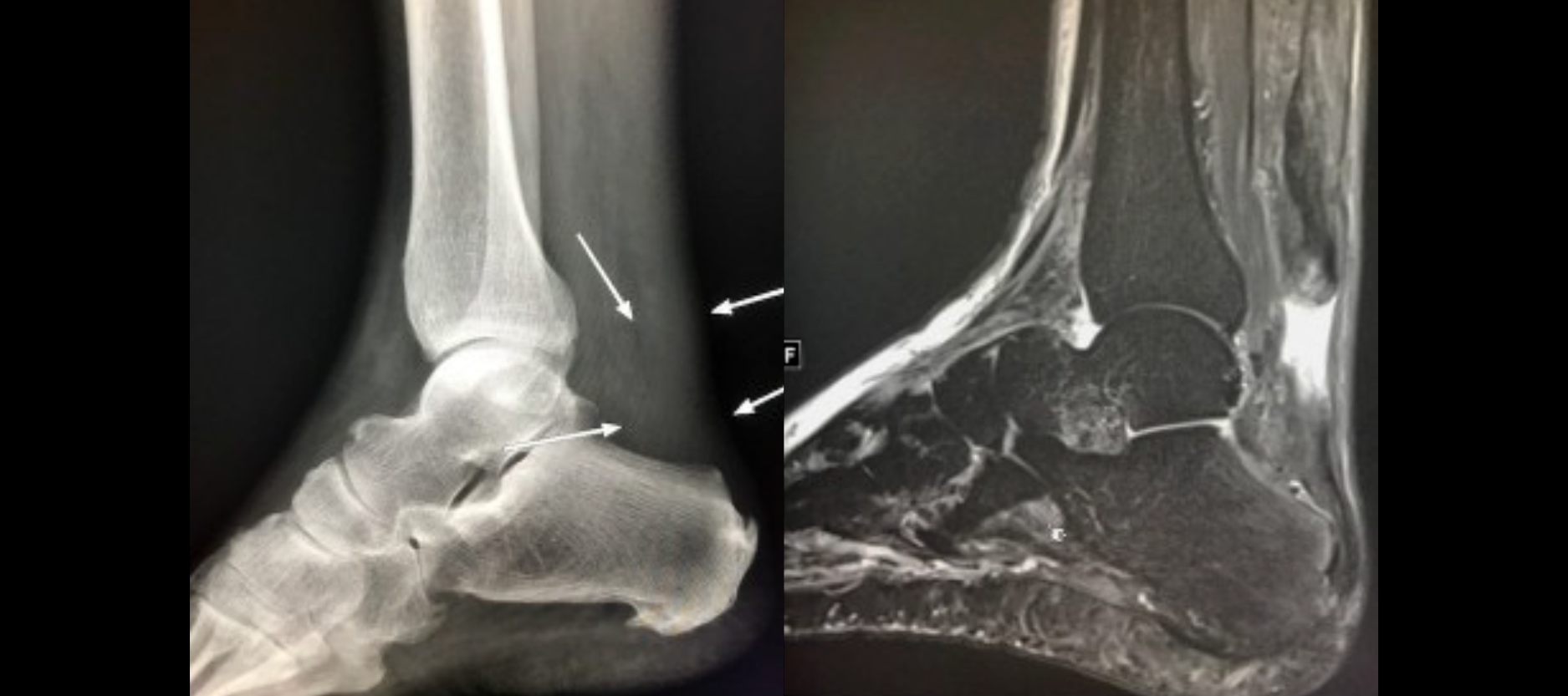
The lateral X-ray of the ankle demonstrates attenuation in Kager’s fat pad with poor definition of the posterior border of Kager’s fat pad which correlates to the Achilles tendon (arrows). There is also poor definition of the fat stripe around the attenuation typically seen in the region of the Achilles tendon (arrows). This is a sign on Xray that underlying Achilles tendon pathology is likely.
The MRI which followed then clearly demonstrates an Achilles tendon tear.
This case therefore clearly demonstrates the importance of closely evaluating and commenting on soft tissues on X-ray.
In this case the soft tissue abnormality not seen by the referring clinician, but seen and reported by the radiologist, allowed the patient to get the MRI performed in a timely fashion and then appropriately treated by a specialist.