Jonathan S. Luchs, MD, FACR
Chief Medical Officer, Premier Radiology Services
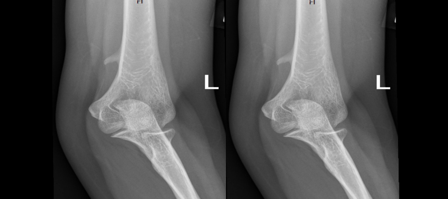
Avian Spur: Also known as supracondylar spur, supracondylar process, supratrochlear spur of the distal anteromedial humeral cortex
Anatomical variant ~1% of the population.
Typically located:
- Anteromedial humeral cortex
- 5 cm proximal to medial epicondyle
- Pointing toward the joint and medial epicondyle of the humerus (as opposed to osteochondroma, which points away from the joint)
This structure is often considered a vestigial structure, as a supracondylar canal (or foramen) can be found as a normal structure in many more primitive mammals, such as felines.
The ligament of Struthers may be present and join the tip of this process with the medial epicondyle.
- Forms a supracondylar canal through which the median nerve and brachial artery pass
- This ligament is thought to represent the vestigial third head of the coracobrachialis muscle.
Most patients are asymptomatic
However, this should be considered if patients present with symptoms of median nerve compression (supracondylar process syndrome) and there is no pathology on imaging of the carpal tunnel-associated neuropathy of the median nerve.
This condition can also result in compression of the brachial artery.
References
- Andreisek G, Crook D, Burg D, Marincek B, Weishaupt D. Peripheral Neuropathies of the Median, Radial, and Ulnar Nerves: MR Imaging Features. Radiographics. 2006;26(5):1267-87. doi:10.1148/rg.265055712– Pubmed
- Ross, Lawrence M., Lamperti, Edward D., Schumacher, Udo et al. Thieme Atlas of Anatomy: General Anatomy and Musculoskeletal System. (2010) ISBN: 1604062924 – Google Books
- Stephen M. Russell. Examination of Peripheral Nerve Injuries: An Anatomical Approach. (2006) ISBN: 1588904830 – Google Books
- Last, R. J., McMinn, R. M. H.. Last’s Anatomy, Regional and Applied. (1994) ISBN: 044304662X – Google Books
- https://radiopaedia.org/articles/supracondylar-spur