Jonathan S. Luchs, MD, FACR
Chief Medical Officer, Premier Radiology Services
Named for Jacques Lisfranc of St. Martin France (1790-1847), a field surgeon in Napoleon’s army serving on the Russian front
Lisfranc joint = the articulation between the midfoot and forefoot, is composed of the five tarsometatarsal (TMT) joints.
Lisfranc injury = indicates an injury to the normal alignment of the cuneiforms and metatarsal joints with the loss of their normal spatial relationships.
Most Common: Occurs at the joint involving the 1st and 2nd metatarsals and the medial cuneiform.
Incidence = 0.2% of all fractures
INJURY MECHANISM: (varied)
- Direct load/crush injury
- Indirect load onto plantar flexed foot
- Tarsometatarsal dislocation may also occur in the diabetic neuropathic joint (Charcot Joints)
SUBTYPES – Lisfranc Fracture Dislocation:
- homolateral: a homolateral injury is a lateral displacement of the 1st to 5th metatarsals, or of 2nd to 5th metatarsals where the 1st MTP joint remains congruent
- divergent: a divergent injury is a lateral dislocation of the 2nd to 5th metatarsals with medial dislocation of the 1st metatarsal
- isolated: this involves one or two metatarsals that dislocate dorsally in isolation
IMAGING:
Most well demonstrated on the standard X-ray views of the foot
Need High Index of Suspicion
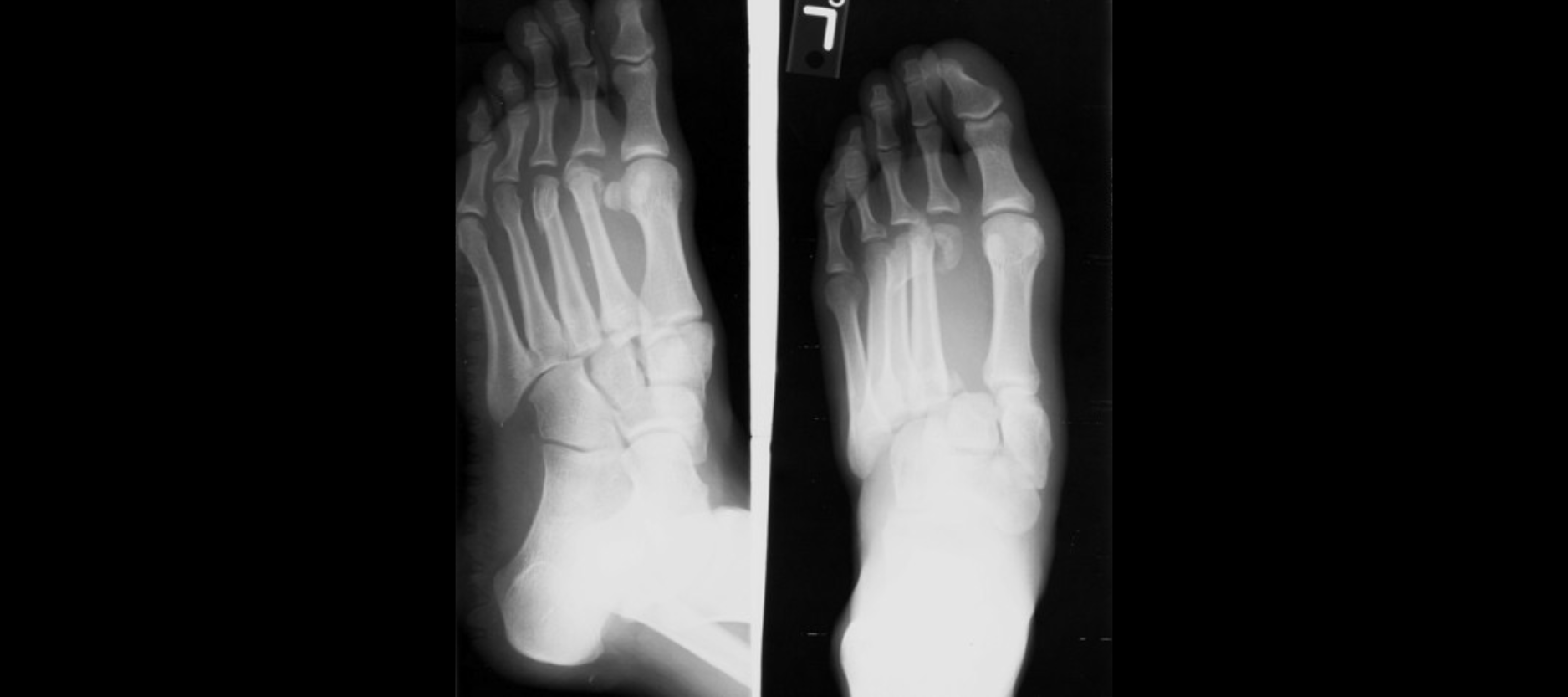
AP View:
Loss normal alignment medial border 2nd MT to Middle Cuneiform
Diastasis between 1st and 2nd MT >2.7mm
FLECK SIGN = small bony fragment base of 2nd MT or Medial Cuneiform reflecting fracture avulsion Lisfranc Ligament
Oblique View:
Loss Normal Alignment 2nd-4th TMT Joints
Lateral View:
Dorsal Displacement base 2nd MT
Flattening Longitudinal Arch
Dorsal Soft Tissue Swelling at TMT Region
Advanced X-ray = stress views with forefoot abduction.
CT– will also demonstrate unsuspected associated fractures.
Associated fractures most often occur at the base of the second metatarsal. They may also be seen in the 3rd metatarsal, 1st or 2nd cuneiform, or navicular bones.
MRI – Assess ligamentous injury if high clinical concern when routine x-rays are inconclusive
TREATMENT AND PROGNOSIS:
Internal fixation = most common treatment.
COMPLICATIONS:
Most common = non-union and post-traumatic arthritis.
Conventional radiography can demonstrate these complications however CT is better for demonstrating these details.
Reference Materials: Lisfranc Injury